Authors: Ciara A. Kerney, Kirsten Hodges, ASA and Jeffrey Doyle, ASA

![]() WHAT IS A GAMMA CAMERA?
WHAT IS A GAMMA CAMERA?
Nuclear medicine is a subspecialty within radiology that utilizes small amounts of radioactive material in order to assess bodily function and anatomical structure in the diagnosis and treatment of human disease. Within nuclear medicine, a healthcare professional introduces – usually via injection – a small amount of radiopharmaceutical called a ‘tracer’ or ‘radiotracer’ into a patient and, subsequently, utilizes a nuclear camera (i.e., a gamma camera) to produce three-dimensional (“3D”) images. Gamma cameras are commonly used in the early detection, diagnosis and treatment planning of various medical ailments including, but not limited to, heart disease, internal bleeding, brain disorders (e.g., Parkinson’s disease), and cancerous tumors.
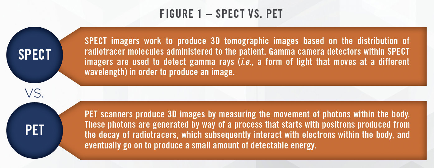
While nuclear medicine is made up of various imaging modalities, the two most commonly deployed are single photon emission computed tomography (“SPECT”) and positron emission tomography (“PET”) scans. The specific radiotracer administered to the patient determines the type of scan the patient will receive (e.g., SPECT or PET). FIGURE 1 provides a brief overview of the SPECT vs. PET imaging modalities.

![]() HISTORY OF NUCLEAR MEDICINE
HISTORY OF NUCLEAR MEDICINE
With roots in the contributions of professionals within the fields of physics, chemistry, and engineering, nuclear medicine was first recognized as an up-and-coming medical specialty in 1946 when it was described in the Journal of the American Medical Association in a report on the success of using radioactive iodine in thyroid cancer treatment.[1] In 1957, American biophysicist and electrical engineer Hal Anger developed the first successful radioisotope camera, or gamma camera, often referred to as the “Anger” camera. The creation of the Anger camera, still utilized today, served as the spark that pushed nuclear medicine out of the laboratory setting and into its current role as an essential part of modern medicine. With the use of nuclear medicine and gamma cameras growing in prevalence within the clinical setting, the American Medical Association (i.e., AMA) officially acknowledged nuclear medicine as an official medical specialty in 1971.
![]() COMBINED TECHNOLOGY
COMBINED TECHNOLOGY
Nuclear imaging technology in its most current form exists as a result of the accumulation of the functions of various singular imaging modalities (e.g., SPECT, PET, CT, MRI) into numerous multifaceted, two-in-one imaging machines. While nuclear imaging started with SPECT and PET, the advanced medical imaging devices outlined below are representative of the consistent innovation within nuclear medicine, all toward the ultimate goal of increased diagnostic accuracy and improved patient outcomes.
PET / CT
Positron emission tomography-computed tomography (i.e., PET/CT) is a medical imaging technique that combines PET and CT technologies to provide simultaneous images of the body’s internal functions using radiotracers (PET) and cross-sectional x-rays to view anatomical structures (CT). Combining the benefits of CT with those of PET generates more comprehensive PET/CT images with a higher degree of accuracy. The accuracy and detail in these images facilitate advanced diagnosis and treatment planning, particularly in areas such as cardiology, neurology, and oncology. Read more about CT technologies in HealthCare Appraisers’ recent article.
PET / MRI
Positron emission tomography (i.e., PET) technology is combined with magnetic resonance imaging (i.e., MRI) to provide a complete ‘picture’ of the body’s processes and structures through the use of PET/MRI. A PET/MRI system uses radiotracers to visualize metabolic processes within the body (PET) and strong magnetic fields to produce detailed images of organs, tissues, and blood vessels (MRI). Similar to the PET/CT, the PET/MRI is used in diagnosis and treatment planning. As further discussed in HealthCare Appraisers’ recent MRI article, MRI machines do not produce radiation, so when compared to a PET/ CT scanner, PET/MRI has a much lower risk of radiation exposure to patients and is, therefore, often used over PET/CT to limit exposure for patients requiring repeated scans. PET/MRI allows for improved motion correction and better soft tissue contrast in comparison to PET/CT, and has applications in the detection of necrotic versus viable tissue after surgical procedures and radiation.
SPECT / CT
Single-photon emission computed tomography (i.e., SPECT/CT) is a hybrid imaging device that provides both functional and anatomical information within a single scan. Generally speaking, SPECT radiotracers tend to be more widely available and have a longer half-life than PET tracers. SPECT/CT systems also have lower spatial resolution than PET/CT, and are, therefore, better suited for functions including imaging blood flow in the heart (i.e., myocardial perfusion imaging), evaluating bone disease, and assessing brain function.
The opportunity for the future development of more highly specialized, cross-functional nuclear imaging devices is seemingly limitless. Experts continue to enter into new territory within nuclear imaging with the development of imaging devices that help fill the gap in services available for the diagnosis of decidedly specific disease, as is the case with the breast specific gamma imaging (i.e., BSGI) technology discussed in HealthCare Appraisers’ recent Mammography article.
![]() APPRAISAL CONSIDERATIONS
APPRAISAL CONSIDERATIONS
![]() Normal Useful Life (NUL) – The NUL considered for a gamma camera under a cost approach to value is estimated to be 10 years. Within a cost approach, an appraiser must consider the maintenance history of the subject machine and assign consideration to software and/ or hardware overhauls and updates. While such updates can be costly, they can significantly extend the NUL of a gamma camera.
Normal Useful Life (NUL) – The NUL considered for a gamma camera under a cost approach to value is estimated to be 10 years. Within a cost approach, an appraiser must consider the maintenance history of the subject machine and assign consideration to software and/ or hardware overhauls and updates. While such updates can be costly, they can significantly extend the NUL of a gamma camera.
![]()
![]()
![]()
![]()
![]()
![]()
![]()
![]()
![]()
![]()
![]()
![]()
![]()
![]()
![]()
![]()
![]()
![]()
![]()
![]()
![]()
![]()
![]()
![]()
![]() CONCLUSION
CONCLUSION
Gamma cameras continue to accelerate the rate of early disease detection, significantly improving treatment options and prognosis by detecting abnormalities and systemic disease processes very early in the progression of disease. As the value of a gamma camera can be heavily dependent upon the considerations discussed herein, it is crucial to engage a qualified and experienced expert within the field of medical equipment appraisal. Healthcare Appraisers’ dedicated team of capital asset appraisers has broad experience in the valuation of gamma cameras and how – among others – the factors above can impact the value of these types of equipment. Contact us today to speak with an expert.
![]() Read the other articles in our Insights on Imaging series:
Read the other articles in our Insights on Imaging series:
[1] https://www.news-medical.net/health/History-of-Nuclear-Medicine.aspx (Accessed August 11, 2023).
[2] Defined by the American Society of Appraisers as “a form of depreciation in which the loss in value is due to factors inherent in the property itself and changes in design, materials, or process that result in inadequacy, overcapacity, excess construction, lack of functional utility, excess operating costs, etc.”
[3] Defined by the American Society of Appraisers as “a form of depreciation or loss in value caused by unfavorable external conditions.”
